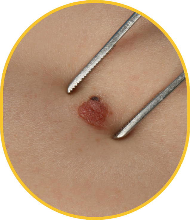

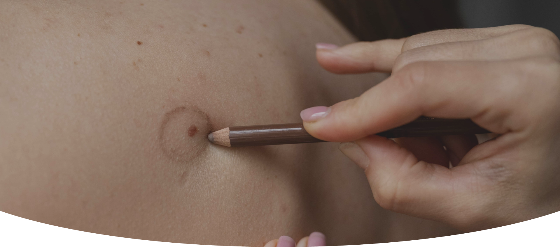
A cherry angioma is a benign vascular lesion formed by the dilation of superficial capillaries in the dermis. It appears as a small, bright red, dome-shaped spot measuring between 1 and 10 mm. Its colour remains stable and does not blanch under pressure, which distinguishes it from other skin lesions. It most often appears on the trunk, neck and arms. A professional assessment confirms the diagnosis and rules out any atypical presentation.

The appearance of cherry angiomas is multifactorial. Skin aging is the main factor: the risk increases significantly after age 40. Genetic predisposition plays an important role, as do hormonal fluctuations related to pregnancy or menopause. Repeated sun exposure, skin trauma, liver insufficiency and certain stimulants, such as coffee, alcohol or tobacco, can also contribute. An increasing number of lesions with age is normal and does not indicate an underlying condition.
A cherry angioma is a strictly benign lesion, with no risk of malignant transformation. It differs structurally and visually from melanoma: its ruby-red colour remains stable, it does not blanch under pressure and its borders remain regular. These criteria clearly distinguish it from suspicious pigmented lesions. Any new skin lesion nevertheless deserves a professional assessment to rule out an atypical presentation. Self-diagnosis carries risks. An in-clinic consultation provides a rigorous differential diagnosis and reassurance based on a clinical examination.
Removing a cherry angioma is a purely elective decision. No medical indication justifies treating a stable benign lesion. Removal becomes relevant when the lesion is located in a visible area such as the face, neck, décolletage or hands, when it bleeds on contact with clothing or while shaving, or when several lesions together create a real aesthetic concern. If a lesion changes in appearance, a professional assessment is recommended before any treatment decision.
Two non-surgical treatments are offered at Clinique Main d'Or to remove cherry angiomas. IPL photorejuvenation targets the vascular lesion through the absorption of intense pulsed light, which causes the visible red spot to coagulate and be eliminated. Electrocoagulation works by applying an electric current targeting the dilated capillary, precisely coagulating it. Our clinic’s professionals determine the most appropriate method during the initial consultation.
Immediately after the session, slight redness or a temporary bluish mark (purpura) may appear for a few days. Small scabs may form and fall off naturally within 7 to 10 days without being picked. Sun protection of the treated area is recommended during healing. For small, isolated angiomas, one to two sessions are generally enough. Results on the treated lesions are long-lasting. However, new angiomas may appear over time, depending on individual predisposing factors.
At Clinique Main d'Or, every treatment begins with a personalized assessment of the skin lesions. Our clinic’s professionals determine the most suitable treatment method based on the type, size and location of the angioma. To understand all the available options, see our pages dedicated to [IPL photorejuvenation] and [electrocoagulation]. The medical-aesthetic clinic is located in Saint-Laurent, Montreal, and supports each person with a personalized protocol.